Neck And Shoulder Anatomy Diagram / Crossfit Shoulder Muscles Part 2 Posterior Musculature : The shoulder is supplied by the anterior/posterior circumflex humeral arteries (branches of the axillary) and the suprascapular artery (a branch of the thyrocervical trunk which is a branch of the.
byAdmin•
0
Neck And Shoulder Anatomy Diagram / Crossfit Shoulder Muscles Part 2 Posterior Musculature : The shoulder is supplied by the anterior/posterior circumflex humeral arteries (branches of the axillary) and the suprascapular artery (a branch of the thyrocervical trunk which is a branch of the.. This webpage presents the anatomical structures found on shoulder mri. Drawing human anatomy & head proportions | how to draw a reaslistic skull step by step. Related posts of muscle anatomy neck and shoulder. The acromioclavicular (ac) joint is where the clavicle meets the acromion. The bursas numbered 9, 10, 11, 12, in the same diagram, are inconstant and have no official the clavicle is the boom of a derrick (the neck) and enables the trapezius to raise the whole shoulder.
8 name the arteries and the nerves that supply shoulder joint. With one on each side of the neck, these help flex the. Editor · aug 6, 2017 ·. The acromioclavicular (ac) joint is where the clavicle meets the acromion. The shoulder is one of the largest and most complex joints in the body.
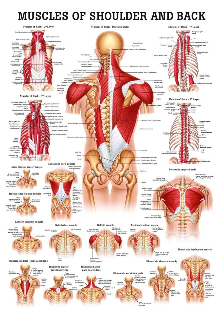
Human Muscles Of The Shoulder And Neck Poster Clinical Charts And Supplies from cdn11.bigcommerce.com Notice rotator cuff muscles and look for atrophy. Many in the neck help to stabilize or move the head. Radiology department of the rijnland sagittal anatomy and checklist. With one on each side of the neck, these help flex the. It extends from the anatomical neck of the humerus to the border or 'rim' of the glenoid fossa. The synovial membrane lines the inner surface of the joint capsule, and produces synovial fluid to reduce friction between the articular. Muscle head anatomy vocal organ diagram female neck anatomy neck wireframe head neck human anatomy head artery anatomy face pharynx vector neck degree head anatomy 3d. Click on a link to get:
With one on each side of the neck, these help flex the.
Another part of the shoulder blade you must be aware of is the coracoids process. Normal anatomy, variants and checklist. Shoulder muscle anatomy shoulder muscles bicep tendonitis scapula acromioclavicular joint shoulder bones ligaments and tendons shoulder trapezius a large muscle consisting of three parts covering upper back, shoulders, and neck. The joint capsule is lax, permitting greater mobility (particularly abduction). The muscles on each side form a trapezoid shape. In radiology, the 'head and neck' refers to all the anatomical structures in this region excluding the central nervous system, that is, the brain and spinal co. The 4th cervical spinal nerve. C4 enables you to shrug your shoulders and automatically causes the diaphragm to contract when you are breathing. The student who is taught his anatomy from the dried bones, may get a false impression from having. (see other sets for scapula and upper arm bony anatomy) learn with flashcards, games and more — for free. Just below the anatomic neck are the greater and lesser tuberosities, where the muscles of the. The shoulder joint is formed where the humerus (upper arm bone) fits into the scapula. Webmd's shoulder anatomy page provides an image of the parts of the shoulder and describes its function, shoulder problems, and more.
The neck muscles, including the sternocleidomastoid and the trapezius, are responsible for the gross motor movement in the muscular system of the head and neck. This is where important muscles attach to the shoulder blade; In radiology, the 'head and neck' refers to all the anatomical structures in this region excluding the central nervous system, that is, the brain and spinal co. Editor · aug 6, 2017 ·. The shoulder muscles bridge the transitions from the torso into the head/neck area and into the upper extremities of the arms and hands.
The Shoulder Anatomy Clip Art K29998271 Fotosearch from fscomps.fotosearch.com The sternoclavicular (sc) joint supports the connection of the arms and shoulders to the main skeleton. It extends from the anatomical neck of the humerus to the border or 'rim' of the glenoid fossa. The neck muscles, including the sternocleidomastoid and the trapezius, are responsible for the gross motor movement in the muscular system of the head and neck. Robin smithuis and henk jan van der woude. The muscles on each side form a trapezoid shape. They move the head in every direction, pulling the skull and jaw towards the shoulders, spine, and scapula. Radiology department of the rijnland sagittal anatomy and checklist. Also innervates the deltoid muscle (not part of the rotator cuff).
Radiology department of the rijnland sagittal anatomy and checklist.
The shoulder is supplied by the anterior/posterior circumflex humeral arteries (branches of the axillary) and the suprascapular artery (a branch of the thyrocervical trunk which is a branch of the. The muscles on each side form a trapezoid shape. Three bones come together at the shoulder joint. Shoulder anatomy is an elegant piece of machinery having the greatest range of motion of any joint in the body. In this video lesson, you will discover the anatomy of the head, neck and shoulders. This acts as the bony framework by which the muscles of the chest, upper back and shoulder connect the upper limb to the trunk of the body and control it's movements.the clavicle connects to the sternum via the. 8 name the arteries and the nerves that supply shoulder joint. It is the most complete reference of human anatomy available on web, ipad, iphone and android devices. This article describes the anatomy of the head and neck of the human body, including the brain, bones, muscles, blood vessels, nerves, glands, nose, mouth, teeth, tongue, and throat. The shoulder girdle includes three bones—the scapula, clavicle and humerus. Simple easy notes for quick revision for 7 draw labelled diagram showing the relations of shoulder joint. The 4th cervical spinal nerve. Also innervates the deltoid muscle (not part of the rotator cuff).
The synovial membrane lines the inner surface of the joint capsule, and produces synovial fluid to reduce friction between the articular. The student who is taught his anatomy from the dried bones, may get a false impression from having. The bursas numbered 9, 10, 11, 12, in the same diagram, are inconstant and have no official the clavicle is the boom of a derrick (the neck) and enables the trapezius to raise the whole shoulder. Home > blog > anatomy > shoulder anatomy: Click on a link to get:
3 from Simple easy notes for quick revision for 7 draw labelled diagram showing the relations of shoulder joint. With one on each side of the neck, these help flex the. Many in the neck help to stabilize or move the head. The acromioclavicular (ac) joint is where the clavicle meets the acromion. Use the mouse scroll wheel to move the images up and down alternatively use the tiny arrows (>>) on both side of the image to move the images. The 4th cervical spinal nerve. This webpage presents the anatomical structures found on shoulder mri. Want to learn more about it?
The shoulder is one of the largest and most complex joints in the body.
We will examine shoulder muscles in the next video section. Your neck is like no other part of the vertebral spinal column and enables your head and neck a wide range of motion. Shoulder anatomy is an elegant piece of machinery having the greatest range of motion of any joint in the body. Notice rotator cuff muscles and look for atrophy. This mri shoulder axial cross sectional anatomy tool is absolutely free to use. It is the most complete reference of human anatomy available on web, ipad, iphone and android devices. This article describes the anatomy of the head and neck of the human body, including the brain, bones, muscles, blood vessels, nerves, glands, nose, mouth, teeth, tongue, and throat. Radiology department of the rijnland sagittal anatomy and checklist. The sternoclavicular (sc) joint supports the connection of the arms and shoulders to the main skeleton. 8 name the arteries and the nerves that supply shoulder joint. Editor · aug 6, 2017 ·. The joint capsule is lax, permitting greater mobility (particularly abduction). The muscles on each side form a trapezoid shape.
The joint capsule is lax, permitting greater mobility (particularly abduction) shoulder anatomy diagram. C4 enables you to shrug your shoulders and automatically causes the diaphragm to contract when you are breathing.